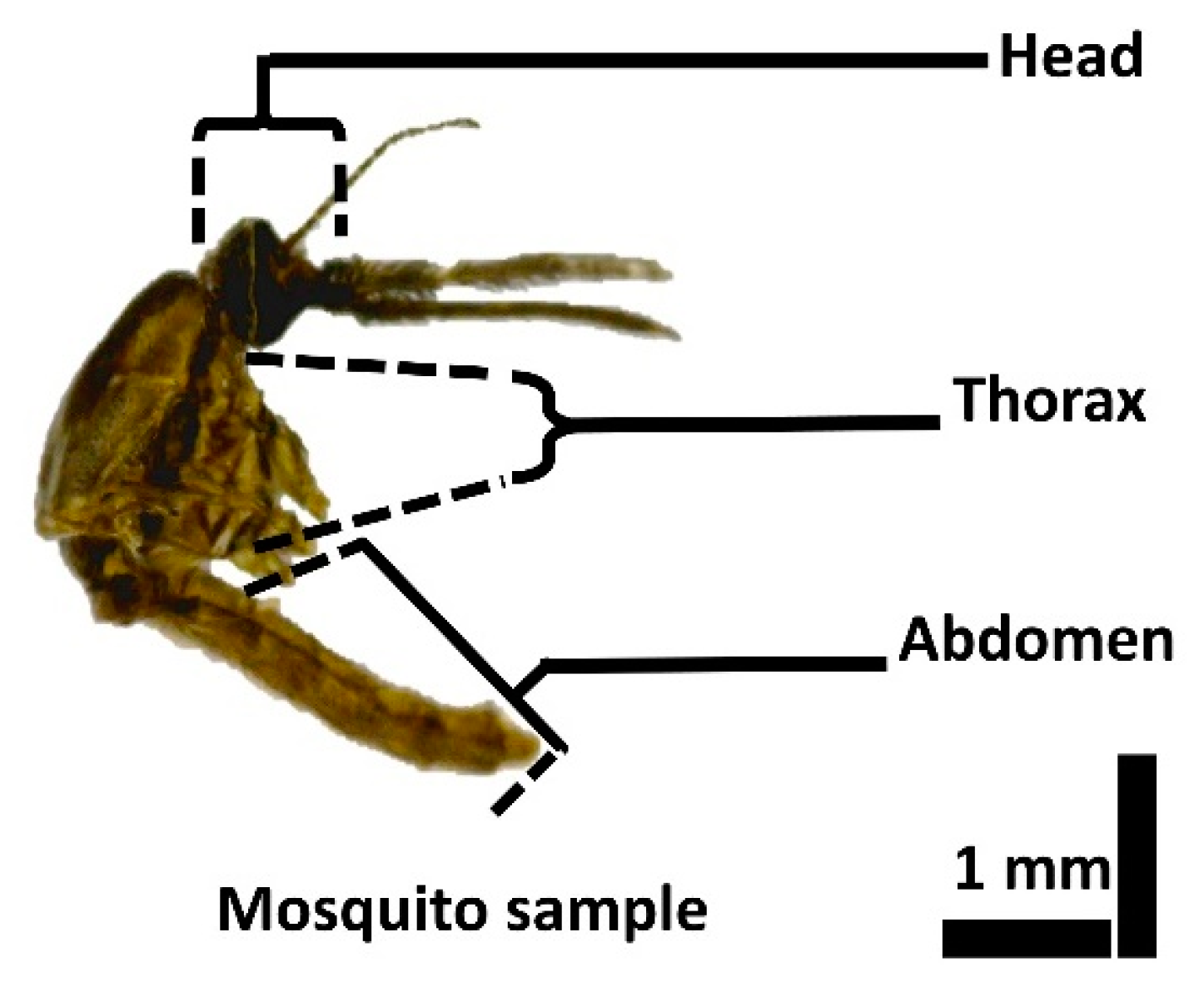
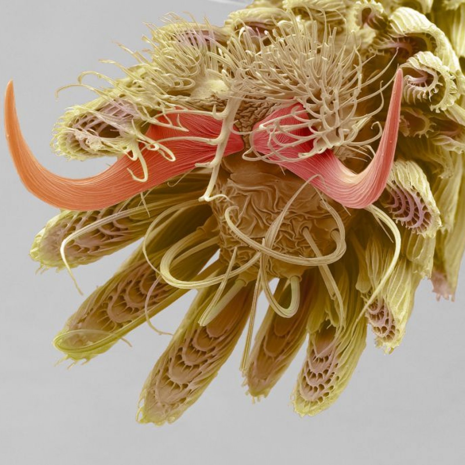
The mouth is made up of two large upper mandibles two lower mandibles known as maxilla the upper lip labrum as well as the lower lip labium. The picture is a mosquitos foot at 800x magnification.
 Small Animals Seen In The Scanning Electron Microscope Scanning Electron Microscope Electron Microscope Microscopic
Superresolution imaging to better understand herpes simplex virus-1 HSV-1.

Mosquito light microscope. Allard From the Department of Pathology Laval University Medical School Quebec Canada and the Institute of Cardiology Maisonneuve Hospital Montreal Canada. Capturing clear images of insects can always be a challenge. The optical microscope also referred to as a light microscope is a type of microscope that commonly uses visible light and a system of lenses to generate magnified images of small objects.
AOMEKIE Stereo Microscope for Kids Students 20-40X with 10Pcs Slides Insect Specimen LED Light Source Wide Field Eyepiece 43 out of 5 stars 50 5299 52. 3D Bug Specimen Kit 5. A stereo dissecting.
Four sides of a mosquito head imaged using a ZEISS light sheet microscope and rendered in Imaris. While you may be aware of the many parts of an eye that some insects have youve probably never seen them on a microscopic level. Optical microscopes are the oldest design of microscope and were possibly invented in their present compound form in the 17th century.
Bodies must be dissected under microscopes. Imaged using an FEI Quanta microscope this frontal view has a real-life width of just 284µm. Chemical analyses must be performed.
Using Dino-Lite microscope cameras and the included software can help make imaging easier and more efficient. A low power stereo microscope disecting microscope is better suited for that purpose. Light optical electron and scanning probe microscopes.
A stereo microscope allows you to see the surface of specimens with a 3-dimensional view. The specimen will appear brightly lit against its contrasting dark field. You can easily modify or adjust the settings of your microscope to illuminate the specimens under a dark field.
Photomicrograph made by Arthur E Smith in the early 1900s using a combined microscope and camera. 3D Bug Specimen Kit 4. Without context that could certainly take some readers by surprise.
Like the bright field microscope a dark field microscope is used in various disciplines such as microbiology and bacteriology. We are able to see in amazing detail the anatomy of the head mouth parts and eyes of the mosquito. 3D Bug Specimen Kit 5 Learn More.
Under the microscope footage of Mosquito Mouth Parts whole mount. On the top is a digital image from a stock QX3 microscope using auxiliary illumination provided by a fiber optic light pipe through a hole drilled into the mixing chamber. An insect becomes infected by biting an infected human being.
The light reflected back out of the crystal. The image on the bottom was recorded using the QX3 microscope body and a Nikon achromatic substage condenser with low numerical aperture. The DinoCapture software allows users to annotate images as well as help organize the images into folders to increase productivity.
The compound eyes and delicate wing structure can be seen like youve never seen it before. If this is the case you will need to dissect the insect and prepare a slide. Light microscopes have a history of more than 500 years.
Observing the microscopic world has never been easier. FEI gives you that opportunity. 3D Bug Specimen Kit 4.
The light microscope also provides a better view of the mouthparts of the ant. In contrast the light has to pass through the specimen to form the image under a compound microscope. Under a stereo microscope you can see the metallic texture and colors of the mosquitos compound eyes.
A light and electron microscope study of the morphological changes induced in rat liver cells by the azo dye 2-me-dab J. Basic optical microscopes can be very simple although many complex. With such a microscope you get a stereoscopic 3D image of the insect.
Stephanie Rainey Maria Vittoria Mancini Colin Loney. You also need to prepare a slide because when using a biological microscope the specimen must be translucent allow light to pass through it. Different light microscopes form a vast family tree and they are still developing.
Atom under the microscope. Depended on the principle of generating images microscopes can be classified into three types. 3D Bug Specimen Kit 4.
They were later published in 1909 in a book called Nature Through Microscope Camera. In 1904 the Royal Society in London exhibited a series of Smiths photomicrographs to the public. This will allow you to view a flat specimen under a biological microscope which has more magnification.
 Mosquito Head High Resolution Stock Photography And Images Alamy
 Anopheles Stephensi Mosquito Female Mosquito Made By Humans Fish Pet
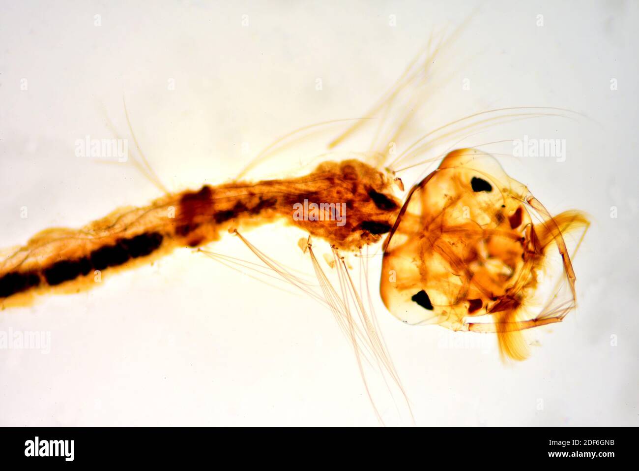
 649 Magnification Mosquito Photos Free Royalty Free Stock Photos From Dreamstime
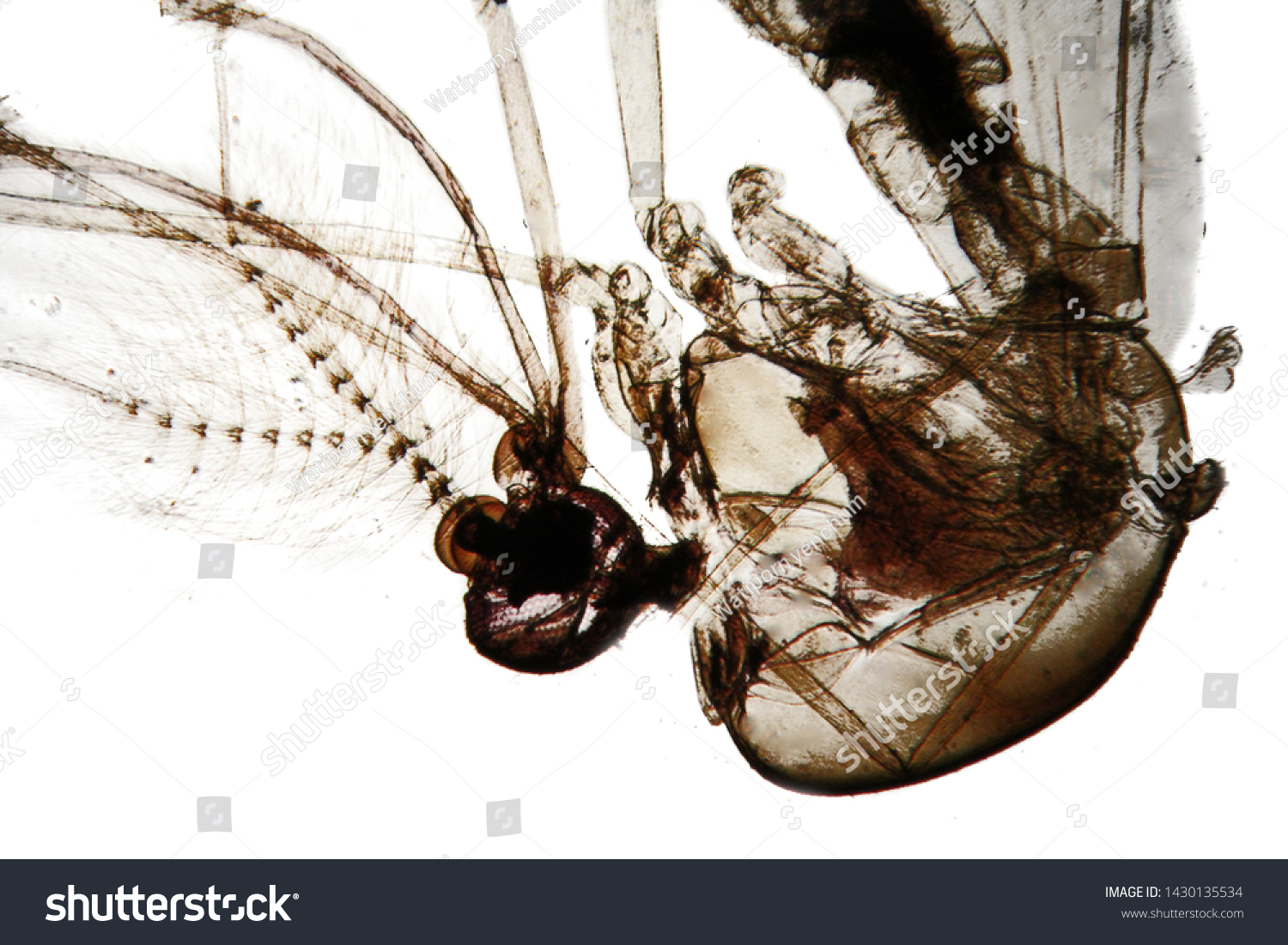 Structure Male Mosquito Show Upper Part Stock Photo Edit Now 1430135534
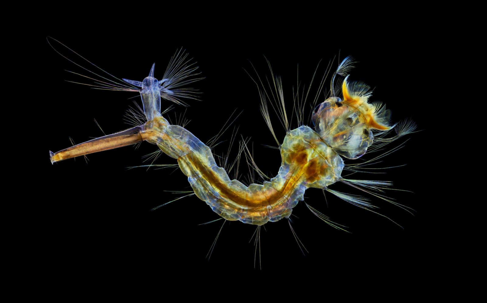 Mosquito Larva 2019 Photomicrography Competition Nikon S Small World
Science Source Stock Photos Video Mosquito Seen Under Microscope Culicidae Lm
 Sensors Free Full Text Non Destructive Analysis Of The Internal Anatomical Structures Of Mosquito Specimens Using Optical Coherence Tomography Html
 Insect Sem Part 3 Microscopic Photography Scanning Electron Microscope Micro Photography
 Mosquito Head Sem By Steve Gschmeissner Mosquito Bugs And Insects Pictures Of Insects
 Mosquito Under Microscope Microscopy
 Scanning Electron Microscopy Sem Images Of A Albopictus Mosquito Download Scientific Diagram
 Light Microscopic Images Of 4 Th Instar Mosquito Larvae Of Culex Download Scientific Diagram
 Under The Microscope Amazing Bizarre Image Of Mosquito Leg Baffles The Internet
 Mosquito Under Microscope Selective Focus Light Micrograph Stock Photo Picture And Royalty Free Image Image 112327482